Largest gland in human body, the liver, is undoubtedly a vital organ and supports every other organ in the body.
The liver
The organs found in every vertebrate (have spines), is the one, which is the largest gland of one of the vertebrate, that is, the human being is, the liver.
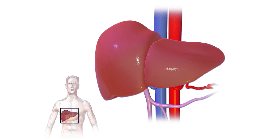
In humans, the liver is located in the upper right quadrant of the abdomen just below the diaphragm. It is a meaty organ weighing around 3 pounds.
Besides, its use as detoxification of metabolites and protein synthesis, it is used as an important digestive organ. Because of the production of cholesterol and bile acids containing, an alkali fluid, named as BileBile, it helps in the breakdown of fats.
In the liver, there is a highly specialized tissue which is made up of main hepatocytes or liver cells.
This tissue regulates a wide variety of highly-rated biochemical reactions which also includes the formation as well as a breakdown of various small and so call Complex molecules.
This biochemical reactions are no doubt always very helpful and necessary for normal functioning of the body and hence proved liver at the largest gland in the human body.
However, besides this, Lever does almost uncountable, or functions and hence is not estimated.
Structure of the liver
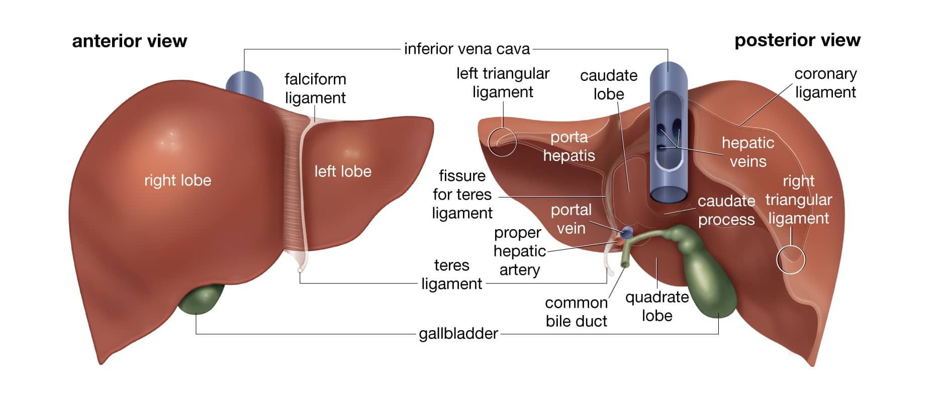
If we talk about the structure of the wedge-shaped, reddish-brown organ, then it must be noted that the liver consists of two unequal sized lobes.
However, the normal weight of a human liver is 3 pound or 1.5 kg, but it where is accordingly human male and female.
In the human male, its size varies between 970 grams to 1850 grams while in the human female, its size varies between 600 grams to 1750 grams.
Thus, focusing on the weight of the liver is also considered as the heaviest inside organ of the human body.
Moving further to the structure of the human liver, it is to be noted that it is connected with two large blood vessels.
That two blood vessels are hepatic artery which carries oxygen-rich blood from an aorta while the other is a portal vein which carries nutrient digested blood from spleen pancreas and gastrointestinal tract.
However, these blood vessels are further divided into capillaries called Liver Sinusoids which later formed the lobules.
These lobules are actually the functional units of the liver.
These hepatocytes formed unit of the liver that is, lobules are actually a functional unit of it.
These lobules are fixed together with a very fine however dense as well as an irregular fibroelastic connective tissue which extends from the fibrous capsule and covers the whole liver which is called as Glisson’s capsule.
Gross anatomy of the liver
When divided by, the following sub organs are there related to the liver which are mentioned below
1.Lobes: when taken our view of the liver, a fine ligament called Falciform ligament divides it into two, that is the right and the left lobe.
While when viewed from below, aside from left and right lobe to other lobes specifically named as Caudate and Quadrate lobes are added to it.
Beside the lobe bifurcation, the gross anatomy also leads to view a line running from the left of the liver.
This line is termed as Cantlie’s line which divided the liver and underneath organ gallbladder into two halves.
However, Lingamentum Venosum an anatomical mark, divides the left part of the liver into two parts.
Meanwhile, Porta Hepatis an important anatomical mark, divides the liver into seven small segments.
2.Surfaces: The Gross Anatomy of the liver deals with the diaphragmatic surface of it.
Under this surface, the liver is covered with a double-layered membrane known as Peritoneium excluding the triangular portion.
This surface covers the convex shape of the two lobes.
However, the peritoneum holds back on itself to form various ligaments.
Nevertheless, the Other surfaces are Visceral and Inferior, which are uneven and concave.
These Visceral and inferior surfaces are also covered with peritoneum which attaches the gallbladder and the porta hepatis.
3.Impression: Gross Anatomy of the liver leaves several Impressions on the surfaces which accommodates with various adjacent structures and organs.
Like below the right lobe and right of the gallbladder, there are two Impressions.
These Impressions are one behind the other.
The impression formed by the hepatic flexure is quite a Shallow colic while the other is deep renal.
In the medium of the renal impression is present, a third impression called a duodenal impression, found between it and the neck of the gallbladder.
While the gastric impression which is moulded over the upper front surface of the stomach and the tuber omental.
Also Read: Longest Bones in the Human Body
Microscopic anatomy of the liver
So, the study of microscopic anatomy or the term Histology makes us aware of the two major types of hepatocytes or liver cells.
The major hepatocytes are Parenchymal cells and Non-Parenchymal cells.
Further talking about the cells, we got to know that the parenchymal cells along constitutes to 70% to 85% of the volume of the liver while the non-parenchymal cells are only 6.5 % of its total volume part. However, it constitutes 40% of its total number.
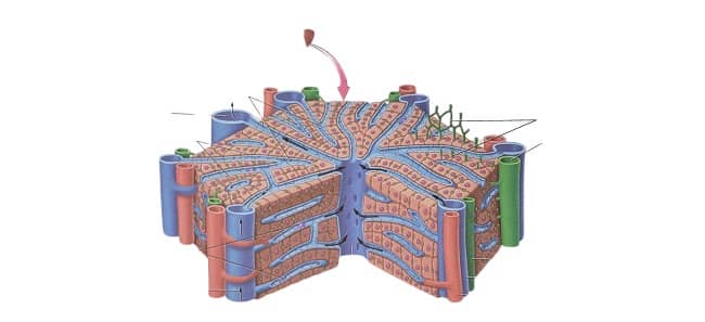
Apart from these, the microscopic anatomy divides the lopes into hepatic lobules.
These lobules are hexagonal in shape and rough enough too.
It consists of plates of hepatocytes and sinusoids radiating from a central vein towards portal triad.
A major function of the central vein is to join the hepatic vein to carry out blood from the liver.
While the portal triad, which consists of the hepatic artery, the portal vein and bile duct seems running along with each lobule’s corners.
Functional anatomy of the liver
Functional anatomy of the liver begins with its Central part known as Hepatic Hilum.
The Hepatic hilum consists of the two openings; the first opening is known as Porta Hepatis. This Porta Hepatis carries Bile duct and Hepatic artery.
While the second opening is for the Portal Vein.
Now, moving forward, these bile ducts, hepatic artery and portal vein gets divided into left and right branches called functional left lobes and right functional lobes respectively.
Now, these functional lobes are further divided or separated by Cantlie’s line.
The plane separates the lobe into true left and true right lobe.
The right lobe is further divided into the anterior and posterior segment by right hepatic vein.
While the left lobe is differentiated into medial and lateral segments by the left hepatic vein.
However, the hepatic hilum is differentiated in terms of three plates, namely Hilar plate, Cystic plate and the umbilical plate.
These plates are the only sights of various anatomical variation.
Functions of the liver
No doubt, the largest gland in the human body, have to go through various functions of the human body.
These functions of the liver is usually done by the hepatocytes.
Some of the important functions done by hepatocytes are listed below:
Blood supply
There are hepatic portal vein as well as hepatic arteries, which makes the liver receives a dual blood supply.
The hepatic portal veins carries the venous blood which are drained from various organs like the spleen, the gastrointestinal tract and various organs.
It delivers around 75% of the blood supply from the liver.
While the hepatic arteries supply arterial blood to the liver.
In this articulation, oxygen demand is fulfilled by both the hepatic portal vein and hepatic arteries.
Biliary flow
According to the name, Biliary flow deals with the flow of bile juice, secreted by the liver.
At first, the BileBile produced in the liver is stored in Bile Canaliculi.
From the Bile canaliculi, the BileBile is transported to the small intestine.
But, it is not necessary that BileBile directly got drained to the duodenum. However, at first, it may be stored to the gall bladder also.
Synthesis
The largest gland in the human body is the site of synthesis as it plays a major role in the synthesis of carbohydrate, protein, amino acid as well as lipid metabolism.
The liver synthesizes and stores glycogen through the process of glycogenesis.
However, when needed to blood, liver secrets glucose to it through the process of glycogenesis.
And through the process of glucogenesis, the synthesis of glucose takes place through amino acids, lactate and glycerol.
The liver is also responsible for protein synthesis, amino acid synthesis, red blood cell production, as well as it plays a great role in clotting factors.
Breakdown
For the breakdown of insulin and hormones, the liver takes place a great role.
Besides this, the liver breaks down bilirubin through the process of glucuronidation and excretes it to BileBile.
However, it plays a key role in the breakdown of waste products, methylation, ammonia as well as drug metabolism.
Blood Reservoir
Being the largest gland in the human body as well as because of its expandable feature, large quantities of blood can be stored in the form of blood vessels.
Because of the high pressure in the right atrium, backpressure is caused in the liver, and it expands to upto 1 litre, and occasionally it got stored in the hepatic veins and sinuses.
Lymph production
The hepatic sinusoids have a high concentration of pores which makes it highly permeable.
With its ability to be highly permeable, the liver sinusoid epithelium allows large quantities of lymph to form.
And hence in the resting position, half of all the lymph is produced in the human body.
Seems a dedicated biology student wrote this
Very well written